|
Overview
 Flat feet- or fallen arches- refers to a condition of the foot in which the arch of the foot collapses while standing (or in some extreme cases, even when no weight is applied to the feet), causing the sole of the foot to become flat. Causes Fallen arches may be caused by a number of causes, including increased elastin during pregnancy, arthritis, injury, excessive stress on the foot, fused bones in the foot, or an extra bone. They may cause not only foot pain, but also pain in the legs, knees, and back and a loss of mobility. The condition is most often treated with orthotics, structures placed in the shoes to support the feet, but this may not be enough for severe cases. Exercises to strengthen and rebuild the arches can also be helpful. Surgery is sometimes the best method of treatment, as it can completely rebuild the arches and has lasting results, but it is quite expensive and considered a last resort. Symptoms Fallen arches symptoms may include the following. Being unable to slip fingers underneath arches. Inwards rolling of foot and ankle when running. Knee problems due to lack of support from feet. Diagnosis If your child has flatfeet, his or her doctor will ask about any family history of flatfeet or inherited foot problems. In a person of any age, the doctor will ask about occupational and recreational activities, previous foot trauma or foot surgery and the type of shoes worn. The doctor will examine your shoes to check for signs of excessive wear. Worn shoes often provide valuable clues to gait problems and poor bone alignment. The doctor will ask you to walk barefoot to evaluate the arches of the feet, to check for out-toeing and to look for other signs of poor foot mechanics. The doctor will examine your feet for foot flexibility and range of motion and feel for any tenderness or bony abnormalities. Depending on the results of this physical examination, foot X-rays may be recommended. X-rays are always performed in a young child with rigid flatfeet and in an adult with acquired flatfeet due to trauma. What is PES Planovalgus deformity? Non Surgical Treatment Most flexible flat feet are asymptomatic, and do not cause pain. In these cases, there is usually no cause for concern, and the condition may be considered a normal human variant. Flat feet were formerly a physical-health reason for service-rejection in many militaries. However, three military studies on asymptomatic adults (see section below), suggest that persons with asymptomatic flat feet are at least as tolerant of foot stress as the population with various grades of arch. Asymptomatic flat feet are no longer a service disqualification in the U.S. military. Surgical Treatment  Procedures may include the following. Fusing foot or ankle bones together (arthrodesis). Removing bones or bony growths, also called spurs (excision). Cutting or changing the shape of the bone (osteotomy). Cleaning the tendons' protective coverings (synovectomy). Adding tendon from other parts of your body to tendons in your foot to help balance the "pull" of the tendons and form an arch (tendon transfer). Grafting bone to your foot to make the arch rise more naturally (lateral column lengthening). Prevention Orthotic inserts, either prescribed or bought over the counter, can help keep the arches fixed into position, but always wear them as although they support, they don?t strengthen, which is why some experts reccomend avoiding them. Gait analysis at a run specialist can help to diagnose overpronation and flat feet. Most brands produce shoes that will give support and help to limit the negative effects of a poor gait on the rest of the body. Barefoot exercises, such as standing on a towel and making fists with the toes, can help to strengthen the arches. Start easy and build up the reps to avoid cramping. Short barefoot running sessions can help take pressure off the arches by using the natural elasticity of the foot?s tendons to take impact and build strength to help prevent flat feet. These should be done on grass for only a few minutes at a time. After Care Patients may go home the day of surgery or they may require an overnight hospital stay. The leg will be placed in a splint or cast and should be kept elevated for the first two weeks. At that point, sutures are removed. A new cast or a removable boot is then placed. It is important that patients do not put any weight on the corrected foot for six to eight weeks following the operation. Patients may begin bearing weight at eight weeks and usually progress to full weightbearing by 10 to 12 weeks. For some patients, weightbearing requires additional time. After 12 weeks, patients commonly can transition to wearing a shoe. Inserts and ankle braces are often used. Physical therapy may be recommended. There are complications that relate to surgery in general. These include the risks associated with anesthesia, infection, damage to nerves and blood vessels, and bleeding or blood clots. Complications following flatfoot surgery may include wound breakdown or nonunion (incomplete healing of the bones). These complications often can be prevented with proper wound care and rehabilitation. Occasionally, patients may notice some discomfort due to prominent hardware. Removal of hardware can be done at a later time if this is an issue. The overall complication rates for flatfoot surgery are low. Overview
Differences between lengths of the two upper extremities (upper and/or lower arms) or between the lengths of the two lower extremities (upper and/or lower legs) are called limb length discrepancy (LLD). A limb length discrepancy may be due to a normal variation that we all have between the two sides of our bodies, or it may be due to other causes. Some differences are so common that they are normal and need no treatment. For example, one study reported that 32 percent of 600 military recruits had a 5mm to 15mm (approximately 1/5 to 3/5 inch) difference between the lengths of their two lower extremities; this is a normal variation. Greater differences may need treatment because a discrepancy can affect a patient?s well being and quality of life. 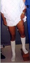 Causes Leg length discrepancies can be caused by: hip and knee replacements, lower limb injuries, bone diseases, neuromuscular issues and congenital problems. Although discrepancies of 2 cm or less are most common, discrepancies can be greater than 6 cm. People who have LLD tend to make up for the difference by over bending their longer leg or standing on the toes of their shorter leg. This compensation leads to an inefficient, up and down gait, which is quite tiring and over time can result in posture problems as well as pain in the back, hips, knees and ankles. Symptoms Many people walk around with LLD?s of up to 2 cm. and not even know it. However, discrepancies above 2 cm. becomes more noticeable, and a slight limp is present. But even up to 3 cm. a small lift compensates very well, and many patients are quite happy with this arrangement. Beyond 3 cm. however, the limp is quite pronounced, and medical care is often sought at that point. Walking with a short leg gait is not only unsightly, but increases energy expenditure during ambulation. It could also put more stress on the long leg, and causes functional scoliosis. Where the discrepancy is more severe, walking becomes grotesque or virtually impossible. Diagnosis A qualified musculoskeletal expert will first take a medical history and conduct a physical exam. Other tests may include X-rays, MRI, or CT scan to diagnose the root cause. Non Surgical Treatment You and your physician should discuss whether treatment is necessary. For minor LLDs in adults with no deformity, treatment may not be necessary. Because the risks may outweigh the benefits, surgical treatment to equalize leg lengths is usually not recommended if the difference is less than one inch. For these small differences, your physician may recommend a shoe lift. A lift fitted to the shoe can often improve your walking and running, as well as relieve back pain caused by LLD. Shoe lifts are inexpensive and can be removed if they are not effective. They do, however, add weight and stiffness to the shoe.  shoe lifts for height Surgical Treatment Shortening techniques can be used after skeletal maturity to achieve leg length equality. Shortening can be done in the proximal femur using a blade plate or hip screw, in the mid-diaphysis of the femur using a closed intramedullary (IM) technique, or in the tibia. Shortening is an accurate technique and involves a much shorter convalescence than lengthening techniques. Quadriceps weakness may occur with femoral shortenings, especially if a mid-diaphyseal shortening of greater than 10% is done. If the femoral shortening is done proximally, no significant weakness should result. Tibial shortening can be done, but there may be a residual bulkiness to the leg, and risks of nonunion and compartment syndrome are higher. If a tibial shortening is done, shortening over an IM nail and prophylactic compartment release are recommended. We limit the use of shortenings to 4 to 5 cm leg length inequality in patients who are skeletally mature. Overview
 Heel pain is most often caused by plantar fasciitis, a condition that is sometimes also called heel spur syndrome when a spur is present. Heel pain may also be due to other causes, such as a stress fracture, tendonitis, arthritis, nerve irritation, or, rarely, a cyst. Because there are several potential causes, it is important to have heel pain properly diagnosed. A foot and ankle surgeon is able to distinguish between all the possibilities and determine the underlying source of your heel pain.What Is Plantar Fasciitis? Plantar fasciitis is an inflammation of the band of tissue (the plantar fascia) that extends from the heel to the toes. In this condition, the fascia first becomes irritated and then inflamed, resulting in heel pain. Causes A flattening or overstretching of your plantar fascia can cause microscopic tears, inflammation, and a burning sensation. While developing slowly, there may be a sudden severe event sometimes occurring in only one foot at a time. Plantar Faciitis can affect people of all ages and backgrounds. Some contributing factors include age, weight-bearing activities, sudden increase in physical activity, improper shoes, excess weight or a recent weight gain (as little as 5 pounds), and poor biomechanics (flat feet, high arches or unnatural gait). Symptoms Pain in the heel can be caused by many things. The commonest cause is plantar fascitis. Other causes include, being overweight, constantly being on your feet, especially on a hard surface like concrete and wearing hard-soled footwear, thinning or weakness of the fat pads of the heel, injury to the bones or padding of the heel, arthritis in the ankle or heel (subtalar) joint, irritation of the nerves on the inner or outer sides of the heel, fracture of the heel bone (calcaneum). Diagnosis The diagnosis of plantar fasciitis is generally made during the history and physical examination. There are several conditions that can cause heel pain, and plantar fasciitis must be distinguished from these conditions. Pain can be referred to the heel and foot from other areas of the body such as the low back, hip, knee, and/or ankle. Special tests to challenge these areas are performed to help confirm the problem is truly coming from the plantar fascia. An X-ray may be ordered to rule out a stress fracture of the heel bone and to see if a bone spur is present that is large enough to cause problems. Other helpful imaging studies include bone scans, MRI, and ultrasound. Ultrasonographic exam may be favored as it is quick, less expensive, and does not expose you to radiation. Laboratory investigation may be necessary in some cases to rule out a systemic illness causing the heel pain, such as rheumatoid arthritis, Reiter's syndrome, or ankylosing spondylitis. These are diseases that affect the entire body but may show up at first as pain in the heel. Non Surgical Treatment Depending on the underlying cause, treatment can include. Rest from activities that stress the heel (such as running and jumping). Ice packs. Regular foot massage, concentrating on the arch of the foot. Professional strapping. A splint worn at night. Flexibility exercises. Ultrasound therapy. Anti-inflammatory medicine (topical or oral). Checking your posture and walking style, to correct imbalances and gait abnormalities that may contribute to the pain. Shoe inserts (orthoses) to help support the foot. In some cases, surgery may be recommended to treat conditions including neuroma, bursitis and heel spurs. Surgical Treatment Extracorporeal shockwave therapy (EST) is a fairly new type of non-invasive treatment. Non-invasive means it does not involve making cuts into your body. EST involves using a device to deliver high-energy soundwaves into your heel. The soundwaves can sometimes cause pain, so a local anaesthetic may be used to numb your heel. It is claimed that EST works in two ways. It is thought to have a "numbing" effect on the nerves that transmit pain signals to your brain, help stimulate and speed up the healing process. However, these claims have not yet been definitively proven. The National Institute for Health and Care Excellence (NICE) has issued guidance about the use of EST for treating plantar fasciitis. NICE states there are no concerns over the safety of EST, but there are uncertainties about how effective the procedure is for treating heel pain. Some studies have reported that EST is more effective than surgery and other non-surgical treatments, while other studies found the procedure to be no better than a placebo (sham treatment). heel bursitis Prevention  You can help to prevent heel pain by maintaining a healthy weight, by warming up before participating in sports and by wearing shoes that support the arch of the foot and cushion the heel. If you are prone to plantar fasciitis, exercises that stretch the Achilles tendon (heel cord) and plantar fascia may help to prevent the area from being injured again. You also can massage the soles of your feet with ice after stressful athletic activities. Sometimes, the only interventions needed are a brief period of rest and new walking or running shoes. Overview
/Sensation---Superficial-Branch-of-Radial---IMG_6132.jpg) A morton's neuroma (or an "inter-digital" neuroma) is found between the toes of the foot, most commonly the third and fourth toes. It can also occur between the metatarsal bones (the long bones in the forefoot). It is basically an entrapped nerve, which becomes inflamed due to constant irritation from the surrounding bony structures. A morton's neuroma (or an "inter-digital" neuroma) is found between the toes of the foot, most commonly the third and fourth toes. It can also occur between the metatarsal bones (the long bones in the forefoot). It is basically an entrapped nerve, which becomes inflamed due to constant irritation from the surrounding bony structures.Causes There are many reasons to develop a neuroma. Improper shoe gear is probably the most likely cause. Repetitive activity and excessive pressure on the ball of the foot are common. Heredity and genetic factors may also be involved. In many cases the structure of the foot may predispose the condition. Associated conditions that may cause neuroma include: bunion, hammer toes, ligament laxity, and/or a tight calf muscle. Some patients may have thinning of the fat pad on the ball of the foot, which may result in increased pressure of the nerves. Tight pointy shoes (and high heels) without padding may induce pain in the ball of the foot. Neuroma may occur suddenly, or develop over time. Symptoms A Morton's neuroma usually causes burning pain, numbness or tingling at the base of the third, fourth or second toes. Pain also can spread from the ball of the foot out to the tips of the toes. In some cases, there also is the sensation of a lump, a fold of sock or a "hot pebble" between the toes. Typically, the pain of a Morton's neuroma is relieved temporarily by taking off your shoes, flexing your toes and rubbing your feet. Symptoms may be aggravated by standing for prolonged periods or by wearing high heels or shoes with a narrow toe box. Diagnosis The most common condition misdiagnosed as Morton's neuroma is metatarsophalangeal (MTP) joint synovitis. When pain occurs in the third interspace, the clinician may misdiagnose the condition as Morton's neuroma instead of MTP synovitis, which may manifest very much like Morton's neuroma. MTP synovitis is distinguished from Morton's neuroma by subtle swelling around the joint, pain localized mainly within the joint, and pain with forced toe flexion. Palpation of the MTP joint is performed best with a pinching maneuver from the dorsal and plantar aspects of the joint to elicit tenderness of the joint. Other conditions often misdiagnosed as Morton's neuroma include the following. Stress fracture of the neck of the metatarsal. Rheumatoid arthritis and other systemic arthritic conditions. Hammertoe. Metatarsalgia (ie, plantar tenderness over the metatarsal head) Less common conditions that have overlapping symptoms with Morton's neuroma include the following. Neoplasms. Metatarsal head osteonecrosis. Freiburg. steochondrosis. Ganglion cysts. Intermetatarsal bursal fluid collections. True neuromas. Non Surgical Treatment The good news is that the pain can often be relieved fairly easily with the right softer styled orthotic (even in those cases where there is concurrent plantar plate tears and capsulitis!), but its important to remeber that even if your no longer in pain, there is no magic cure to speeding up the healing process so one must take care of their feet for 6-12 weeks. As a rule of thumb a neuroma should always be treated conservatively where possible. This means icing and resting the area, trying to remove the causative factors, and providing postural control and support via metatarsal domes or, if needed, specialised pre-made or custom made orthotics.  Surgical Treatment When early treatments fail and the neuroma progresses past the threshold for such options, podiatric surgery may become necessary. The procedure, which removes the inflamed and enlarged nerve, can usually be conducted on an outpatient basis, with a recovery time that is often just a few weeks. Your podiatric physician will thoroughly describe the surgical procedures to be used and the results you can expect. Any pain following surgery is easily managed with medications prescribed by your podiatrist. There are not one but two different types of leg length discrepancies, congenital and acquired. Congenital implies you are born with it. One leg is structurally shorter than the other. As a result of developmental phases of aging, the brain picks up on the gait pattern and identifies some difference. The entire body typically adapts by dipping one shoulder to the "short" side. A difference of less than a quarter inch isn't very irregular, doesn't need Shoe Lifts to compensate and mostly doesn't have a profound effect over a lifetime.
 Leg length inequality goes mainly undiscovered on a daily basis, yet this problem is very easily fixed, and can eliminate a number of incidents of back discomfort. Treatment for leg length inequality usually involves Shoe Lifts . These are typically very reasonably priced, normally costing below twenty dollars, in comparison to a custom orthotic of $200 or higher. Differences over a quarter inch can take their toll on the spine and should probably be compensated for with a heel lift. In some cases, the shortage can be so extreme that it requires a full lift to both the heel and sole of the shoe. Back pain is easily the most common health problem impacting men and women today. Over 80 million people are affected by back pain at some stage in their life. It is a problem which costs companies millions of dollars each year as a result of lost time and output. Fresh and more effective treatment solutions are continually sought after in the hope of minimizing the economical impact this issue causes.  People from all corners of the earth suffer from foot ache as a result of leg length discrepancy. In a lot of these cases Shoe Lifts can be of beneficial. The lifts are capable of relieving any discomfort and pain in the feet. Shoe Lifts are recommended by numerous skilled orthopaedic practitioners". So that you can support the human body in a healthy and balanced manner, your feet have got a significant role to play. In spite of that, it's often the most neglected area in the human body. Some people have flat-feet which means there is unequal force exerted on the feet. This causes other body parts like knees, ankles and backs to be affected too. Shoe Lifts ensure that correct posture and balance are restored. There are actually two different kinds of leg length discrepancies, congenital and acquired. Congenital indicates you are born with it. One leg is structurally shorter compared to the other. As a result of developmental stages of aging, the brain picks up on the walking pattern and identifies some variation. The entire body usually adapts by tilting one shoulder over to the "short" side. A difference of less than a quarter inch is not blatantly irregular, does not need Shoe Lifts to compensate and typically does not have a serious effect over a lifetime.
 Leg length inequality goes largely undiscovered on a daily basis, however this problem is very easily corrected, and can reduce quite a few incidents of back problems. Treatment for leg length inequality usually involves Shoe Lifts. These are affordable, frequently priced at below twenty dollars, in comparison to a custom orthotic of $200 or even more. When the amount of leg length inequality begins to exceed half an inch, a whole sole lift is generally the better choice than a heel lift. This prevents the foot from being unnecessarily stressed in an abnormal position. Upper back pain is easily the most common ailment afflicting men and women today. Around 80 million men and women are affected by back pain at some stage in their life. It is a problem which costs businesses vast amounts of money year after year because of lost time and output. Innovative and better treatment methods are constantly sought after in the hope of decreasing the economic influence this issue causes.  People from all corners of the world suffer the pain of foot ache as a result of leg length discrepancy. In these types of cases Shoe Lifts might be of very helpful. The lifts are capable of alleviating any discomfort and pain in the feet. Shoe Lifts are recommended by numerous qualified orthopaedic physicians. So that you can support the body in a healthy and balanced fashion, your feet have a crucial job to play. Despite that, it's often the most neglected region of the body. Some people have flat-feet meaning there may be unequal force exerted on the feet. This will cause other areas of the body like knees, ankles and backs to be impacted too. Shoe Lifts make sure that the right posture and balance are restored.  Overview The heel bone is the largest bone in the foot and absorbs the most shock and pressure. A heel spur develops as an abnormal growth of the heel bone. Most commonly, calcium deposits form when the plantar fascia pulls away from the heel area, causing a bony protrusion, or heel spur to develop. While bone spurs can develop in other locations such as the rear of the heel bone, this is the most common location for development. The plantar fascia is a broad band of fibrous tissue located along the bottom surface of the foot that runs from the heel to the forefoot. Heel spurs can cause extreme pain in the rearfoot, especially while standing or walking. Causes Early signs of heel pain are usually due to plantar fasciitis, the inflammation of the plantar fascia. It is probably the most common cause of heel pain seen by the podiatrist. It is seen in all groups of people; runners, athletes, weekend warriors, people who have jobs requiring a fair amount of standing, walking, or lifting, and those who have recently gained weight. The pain most often manifests itself after periods of non-weight bearing when the plantar fascia is given a chance to rest, so your first steps cause a sudden strain to the tissue. Pain is most common with the first steps in the morning and after periods of rest. Fortunately, upwards of 80 percent of those treated early do remarkably well with conservative therapy.  Symptoms Pain and discomfort associated with heel spurs does not occur from the spur itself. The bone growth itself has no feeling. However, as you move, this growth digs into sensitive nerves and tissue along the heel of the foot, resulting in severe pain. Pain can also be generated when pushing off with the toes while walking. Swelling along the heel is also common. Diagnosis A thorough history and physical exam is always necessary for the proper diagnosis of heel spurs and other foot conditions. X rays of the heel area are helpful, as excess bone production will be visible. Non Surgical Treatment Podiatric Care for heel spur syndrome may involve keeping the fascia stretched out by performing exercises. Your doctor may also suggest for you to be seen by a physical therapist. You probably will be advised on the best shoes to wear or some inserts for your shoes. Your podiatrist may suggest that a custom made orthotic be made to allow your foot to function in the most ideal way especially if you have excessive pronation. A heel lift may be used if you have a leg length discrepancy. Medical treatment may include anti-inflammatory oral medications or an injection of medication and local anesthetic to reduce the swelling and decrease pain. If a bursitis is present the medication may greatly improve the symptoms. Your podiatric physician may also recommend a surgical procedure to actually fix the structural problem of your foot. Surgical Treatment Usually, heel spurs are curable with conservative treatment. If not, heel spurs are curable with surgery, although there is the possibility of them growing back. About 10% of those who continue to see a physician for plantar fascitis have it for more than a year. If there is limited success after approximately one year of conservative treatment, patients are often advised to have surgery. Prevention Walk around before you buy shoes. Before you purchase your shoes, do the following. Re-lace the shoes if you're trying on athletic shoes. Start at the farthest eyelets and apply even pressure to the laces as you come closer to the tongue of the shoe. Make sure that you can wiggle your toes freely inside of the shoe. Also, make sure that you have at enough space between your tallest toe and the end of the shoe. You should have room equal to about the width of your thumb in the tip of your shoe. Walk around to make sure that the shoe has a firm grip on your heel without sliding up and down. Walk or run a few steps to make sure your shoes are comfortable. Shoes that fit properly require no break-in period.  Overview In the setting of plantar fasciitis, heel spurs are most often seen in middle-aged men and women, but can be found in all age groups. The heel spur itself is not thought to be the primary cause of pain, rather inflammation and irritation of the plantar fascia is thought to be the primary problem. A heel spur diagnosis is made when an x-ray shows a hook of bone protruding from the bottom of the foot at the point where the plantar fascia is attached to the heel bone. Causes A heel spur usually develops as a result of wear and tear over time, which leads to the degeneration of connective tissue called fascia. Standing for prolonged periods and wearing shoes that do not provide the right type of arch support can also lead to connective tissue damage in the heel. The body attempts to repair the damaged tissue by delivering calcium to the affected region, but sometimes too much calcium begins to accumulate and this results in painful plantar fasciitis. 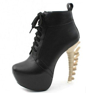 Symptoms The vast majority of people who have heel spurs feel the asscociated pain during their first steps in the morning. The pain is quite intense and felt either the bottom or front of the heel bone. Typically, the sharp pain diminishes after being up for a while but continues as a dull ache. The pain characteristically returns when first standing up after sitting for long periods. Diagnosis Your doctor, when diagnosing and treating this condition will need an x-ray and sometimes a gait analysis to ascertain the exact cause of this condition. If you have pain in the bottom of your foot and you do not have diabetes or a vascular problem, some of the over-the-counter anti-inflammatory products such as Advil or Ibuprofin are helpful in eradicating the pain. Pain creams, such as Neuro-eze, BioFreeze & Boswella Cream can help to relieve pain and help increase circulation. Non Surgical Treatment There are both conservative and surgical heel spur treatment options. Because the heel pain caused by heel spurs is symptomatic of inflammation, the first step is to ice the area in hopes to reduce the inflammation. The next step is to see our orthopedic specialist to prescribe an appropriate treatment plan. Some conservative treatment options might include Anti-inflammatory medications. Shoe orthotics. Shoe inserts. If conservative treatments are not working, surgery may be required to remove the heel spur. As in all cases of heel pain, it is important to see an orthopedic doctor who specializes in foot and ankle pain. Surgical Treatment Heel spur surgery should only be considered after less invasive treatment methods have been explored and ruled insufficient. The traditional surgical approach to treating heel spurs requires a scalpel cut to the bottom of the food which allows the surgeon to access the bone spur. Endoscopic plantar fasciotomies (EPF) involve one or two small incisions in the foot which allow the surgeon to access and operate on the bone spur endoscopically. Taking a surgical approach to heel spur treatment is a topic to explore with a foot and ankle specialist. Prevention Heel spurs and plantar fasciitis can only be prevented by treating any underlying associated inflammatory disease. Overview
Retrocalcaneal and retroachilles bursitis are both common types of bursitis that can cause pain at the back of the foot, just above the heel. This pain, and often swelling, occurs when one or both bursae at the back of the heel become irritated and inflamed. Because they have similar symptoms, heel bursitis is sometimes confused with problems affecting the Achilles tendon, which is a long strip of fibrous tissue that connects the back of the heel to the calf muscle. Other times, Achilles tendon problems may bring about the bursitis. For example, tight calf muscles can contribute to chronic pulling where the Achilles tendon inserts into the back of the heel. This pulling can result in friction and irritation that eventually lead to bursitis. Causes Inflammation of the calcaneal bursae is most commonly caused by repetitive overuse and cumulative trauma, as seen in runners wearing tight-fitting shoes. Such bursitis may also be associated with conditions such as gout, rheumatoid arthritis, and seronegative spondyloarthropathies. In some cases, subtendinous calcaneal bursitis is caused by bursal impingement between the Achilles tendon and an excessively prominent posterior superior aspect of a calcaneus that has been affected by Haglund deformity. Symptoms You might have Retrocalcaneal Bursitis if you notice any of the following symptoms. You have pain or tenderness at the back of the heel where the Achille's tendon attaches. Have swelling near the attachment of the tendon to the heel bone. You have noticed a slowly growing bump on the back of the heel. The back of the heel turns red after getting rubbed in shoes. The back of the heel hurts worse when you run, walk up hill or wear high heels. Diagnosis Medical examination is not necessarily required in light cases where the tenderness is minimal. In all cases where smooth improvement is not experienced, medical attention should be sought as soon as possible to exclude a (partial) rupture of the Achilles tendon or rupture of the soleus muscle. This situation is best determined by use of ultrasound scanning, as a number of injuries requiring treatment can easily be overlooked during a clinical examination (Ultrasonic image). Ultrasound scanning enables an evaluation of the extent of the change in the tendon, inflammation of the tendon (tendinitis), development of cicatricial tissue (tendinosis), calcification, inflammation of the tissue surrounding the tendon (peritendinitis), inflammation of the bursa (bursitis), as well as (partial) rupture. Non Surgical Treatment Relieving the symptoms of bursitis initially focuses on taking the pressure off the bursa. This can be done with proper cushioning, inserts, or footwear but may require surgery if it is a bone formation problem (i.e. Huglund's Deformity). If your bursitis is caused by an infection (septic bursitis), the doctor will probably drain the bursa sac with a needle and prescribe antibiotics to treat the infection. Surgical Treatment Only if non-surgical attempts at treatment fail, will it make sense to consider surgery. Surgery for retrocalcanel bursitis can include many different procedures. Some of these include removal of the bursa, removing any excess bone at the back of the heel (calcaneal exostectomy), and occasionally detachment and re-attachment of the Achilles tendon. If the foot structure and shape of the heel bone is a primary cause of the bursitis, surgery to re-align the heel bone (calcaneal osteotomy) may be considered. Regardless of which exact surgery is planned, the goal is always to decrease pain and correct the deformity. The idea is to get you back to the activities that you really enjoy. Your foot and ankle surgeon will determine the exact surgical procedure that is most likely to correct the problem in your case. But if you have to have surgery, you can work together to develop a plan that will help assure success. Prevention You can avoid the situation all together if you stop activity as soon as you see, and feel, the signs. Many runners attempt to push through pain, but ignoring symptoms only leads to more problems. It?s better to take some time off right away than to end up taking far more time off later. Runners aren?t the only ones at risk. The condition can happen to any type of athlete of any age. For all you women out there who love to wear high-heels-you?re at a greater risk as well. Plus, anyone whose shoes are too tight can end up with calcaneal bursitis, so make sure your footwear fits. If the outside of your heel and ankle hurts, calcaneal bursitis could be to blame. Get it checked out.  Overview OverviewA hammertoes can be flexible or rigid. Hammertoes often start out flexible and become rigid over time as your toe becomes accustomed to its crooked position. Flexible hammertoes are less serious than rigid hammertoes, as they can be easily diagnosed and treated in their initial stages. Flexible hammertoes are named as such because your affected toe still possesses some degree of movement. Causes Hammer toe usually affects the second toe. However, it may also affect the other toes. The toe moves into a claw-like position. The most common cause of hammer toe is wearing short, narrow shoes that are too tight. The toe is forced into a bent position. Muscles and tendons in the toe tighten and become shorter. Hammer toe is more likely to occur in women who wear shoes that do not fit well or have high heels and children who keep wearing shoes they have outgrown. The condition may be present at birth (congenital) or develop over time. In rare cases, all of the toes are affected. This may be caused by a problem with the nerves or spinal cord. .jpg%3Fformat%3D1500w) Symptoms SymptomsThe most obvious symptoms of this injury will be the the middle toe joint is permanently bent at an angle. In the beginning movement may still be possible but as time passes and the injury worsens the toe will be locked in place and possible require hammer toe correction surgery to fix. Another key indicator of hammer toe is that a lump or corn will form on top of the toe. The toe joint will be painful and walking can cause severe discomfort. Occasionally a callus may form on the sole of the injured foot. If you see any of these symptoms together or have been enduring pain for some time, seeing a podiatrist should be your next step. Diagnosis Although hammertoes are readily apparent, to arrive at a diagnosis the foot and ankle surgeon will obtain a thorough history of your symptoms and examine your foot. During the physical examination, the doctor may attempt to reproduce your symptoms by manipulating your foot and will study the contractures of the toes. In addition, the foot and ankle surgeon may take x-rays to determine the degree of the deformities and assess any changes that may have occurred. Non Surgical Treatment Podiatric Care may include using anti-inflammatory oral medications or an injection of medication and local anesthetic to reduce this swelling. When you go to your doctor, x-rays are usually required to evaluate the structure of your foot, check for fractures and determine the cause. The podiatrist may see you to take care of any corns that develop due to the bone deformities. They may advise you on different shoewear or prescribe a custom made orthotic to try and control the foot structure. Padding techniques may be used to straighten the toe if the deformity is flexible, or pads may be used to lessen the pressure on the area of the corn or ulcer. Your podiatric physician may also recommend a surgical procedure to actually fix Hammer toes the structural problem of your foot. Surgical Treatment If you have a severe case of hammer toe or if the affected toe is no longer flexible, you may need surgery to straighten your toe joint. Surgery requires only a local anesthetic (numbing medicine for the affected area) and is usually an outpatient procedure. This means you don?t have to stay in the hospital for the surgery. |
